by: Morgan Fimreite
After taking Genetics with Dr. King and learning that it’s possible to edit a person’s genome in order to help treat a genetic disorder, I knew I wanted to work in gene therapy. I applied to multiple research programs for the summer, and the second one I heard back from was an offer from Dr. Richard Vile at the Mayo Clinic in Rochester, MN. Not only was I going to be working in Gene Therapy, but specifically in Mayo’s Virology & Gene Therapy (VGT) SURF program. I was beyond ecstatic.
Being in a much larger city, surrounded by strangers instead of friends and familiar professors, had a large impact on my anxiety as I was starting this fellowship; however, as I came to know the people I worked with, I realized there was no reason for me to be afraid. After orientation, I was the last person left on the elevator, going to the 18th floor out of 20, but I quickly found a small group of people next to the locked door that led back to the labs. Dr. Schulze, the coordinator of the VGT SURF program, handed me a packet with all the important information I needed to know.
While waiting for the last few students to join us before going through the locked door, my PI (Principal Investigator), the head of the lab, exited the elevator. Dr. Schulze introduced us to each other and let him lead me back into the lab. Dr. Vile introduced me to my mentor, Jason, and I spent most of my first few days observing and taking notes while Jason told me about the project I would be working on. At the beginning of my project, I understood the basic concepts of what I was doing, and I was reading what felt like my weight in scientific papers. After a few weeks, I was comfortable working in the lab, understanding what I was doing and how it related to my project, and Jason was asking for my input when designing future experiments. Before working on this project, I had always followed the instructions or experimental procedure given to me; I had never helped make decisions, so I felt a new sense of responsibility.
My project was focused on infecting Diffuse Midline Glioma (DMG) cells, brain tumors, with a nonpathogenic retrovirus in hopes of it one day becoming a treatment for this cancer. DMGs are usually diagnosed between the ages of five and nine and have a median survival of about one year after diagnosis. With no viable treatment, DMG is a terrifying diagnosis and a nightmare for parents and children. The virus I was working with was a nonpathogenic retrovirus called Foamy Virus because of its tendency to form syncytia, or bubble-like cell fusion. I would be comparing how Foamy Virus infected and killed both non-DMG brain cancer cells and DMGs.
My first task was to genotype the cells to find out whether they had the mutation that characterizes DMGs. This mutation prevents methylation on a histone variant which is thought to cause a more open chromatin phenotype, possibly making it easier for Foamy to infect. We stained the histones in order to visualize the mutation status via Western blotting, and isolated the DNA to observe the single point mutation in the amino acid sequence with Sanger Sequencing. Then, it was time to determine how well Foamy infected the cells. The Foamy we were infecting cells with had green fluorescent protein (GFP) inserted to its genome, so when the virus has infected a cell, it would fluoresce green. I infected cells with different dilutions and MOIs (multiplicities of infection) and assessed how many cells were infected with Fluorescence Activated Cell Sorting (FACS). We also used FACS to determine how many cells were killed by Foamy. With this data about the susceptibility of each of the cell lines to Foamy, we chose a few cell lines to conduct longer infections with at a lower dose of virus as well as cells to overexpress the mutation. I overexpressed both the mutation and wild type in cells via Lentiviral transduction, which means that cells were infected with another retrovirus (Lentivirus) in order to insert the gene of interest, either mutation or wild type, into cells. I selected for cells that had the lentiviral insert with puromycin, which would kill any cells that didn’t have the puromycin-resistant gene that is inserted with the gene of interest. This was one of the most exciting things I did all summer and was something I very clearly remember learning about in Genetics with Dr. King.
During my last week, as I was finishing up experiments, I worked on the final presentation I was going to give to the Vile Lab and the poster I would present at the SURF symposium. Looking at all the data I collected over my fellowship, we found that at higher doses with shorter infections gave relatively the same results across the cell lines. However, when a lower dose infected cells for a longer amount of time, there was more of a difference in both infectivity and killing between the mutation and wild type cells.
I will never forget the overwhelming kindness that was shown to me, not only in the Vile Lab, but from everyone I had the privilege of meeting and working with at the Mayo Clinic. A simple “thank you” to my mentor, Jason Tonne, and the Vile Lab cannot express how much gratitude I have because of this experience and for all of their help and guidance. It is clear that everyone wants you to succeed, even though it may not happen. In science, there will always be challenges and setbacks, but the atmosphere in the Vile Lab is always hopeful.
Maybe it was because DMGs heavily affect pediatric patients, maybe it was the freezer labels of superheros (and Elvis), or the pictures of cells that look like teddy bears or Pacman, but there always seemed to be some sort of magic in the air, similar to what I felt when I was being treated for Leukemia at American Family Children’s Hospital. It reminded me of why I chose to study biology in the first place and helped me decide that I want to pursue finding better treatments for cancer in the future. I hope to visit the Vile Lab again and see how much progress they’ve made, but for now, I’m back to my studies here at SNC and hope everyone has a great fall semester!
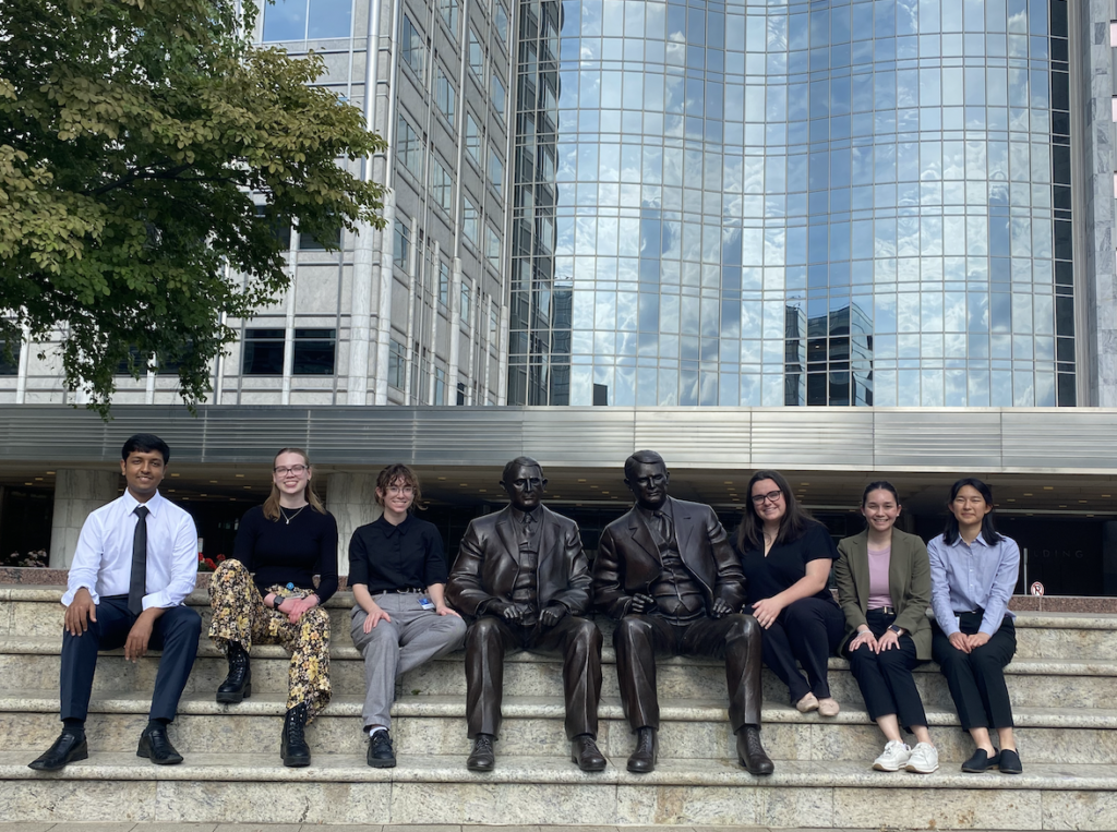
Six of the VGT SURF students posing with the Mayo brothers (from left to right: Showmick, Morgan, Kate, Coryn, Kristin, and Amy).
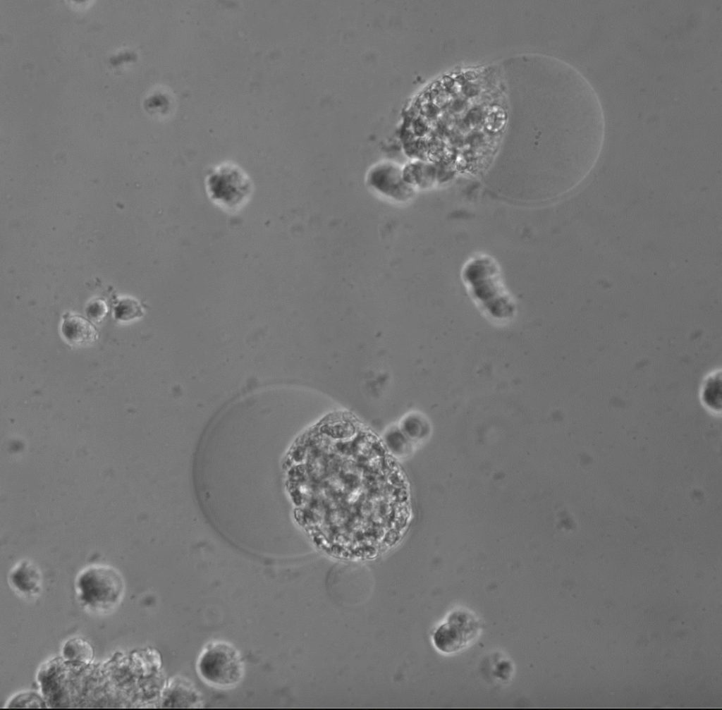
DMG cells infected with Foamy exhibiting syncytia.
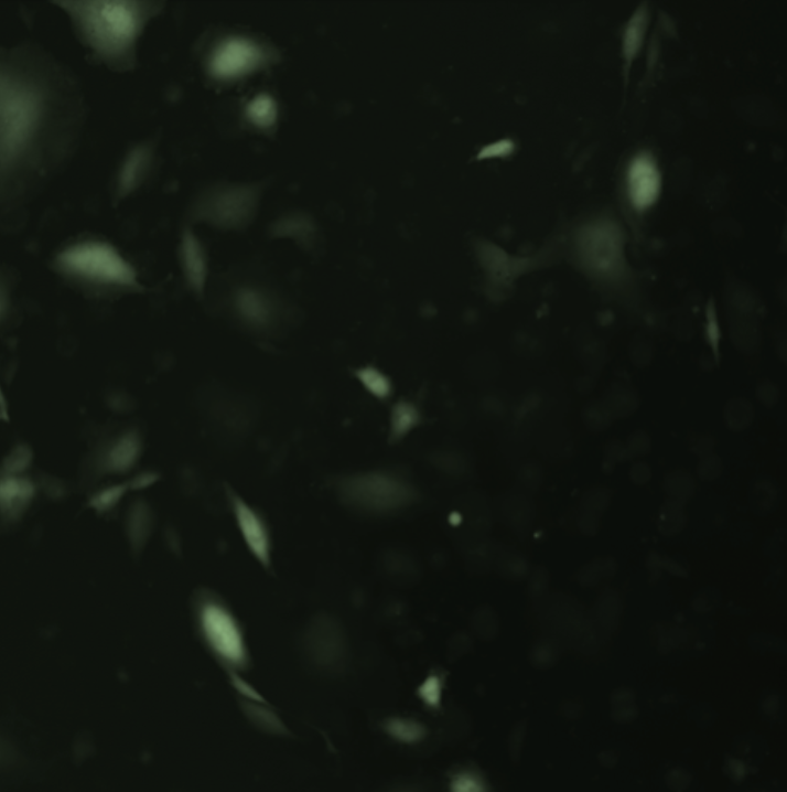
Glioblastoma cells infected with Foamy viewed under fluorescence to view GFP.
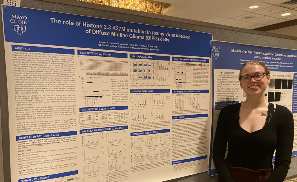
Morgan next to her poster at the Mayo Clinic SURF Symposium.
Morgan what amazing work you are doing. We are so proud of you.